


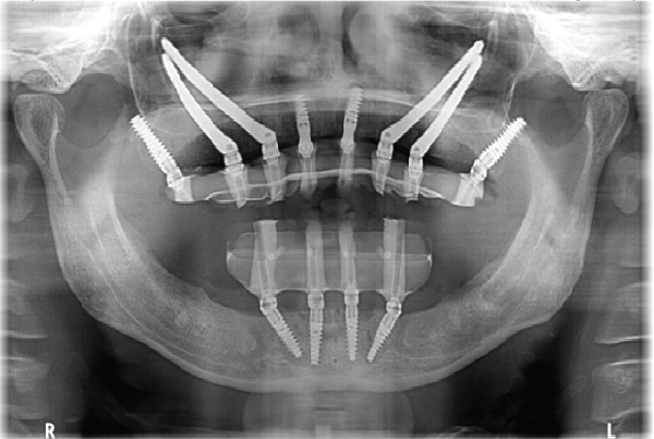
You visit a dentist because one tooth hurts. The dentist examines your mouth, but there may be problems hiding below the gum line or inside the jaw that cannot be seen during a normal visual examination.
This is where dental X-rays can provide useful information. A full-mouth imaging assessment can help a dentist understand the condition of your teeth, roots, jawbone, and surrounding structures before recommending treatment.
Looking at your teeth in a mirror can only show what is visible.
Cavities between teeth, impacted teeth, changes around tooth roots, bone loss, and other conditions may require dental imaging to assess properly.
Dental X-rays are not something you necessarily need at every appointment. The type and frequency of imaging should depend on your dental history, symptoms, examination findings, age, risk factors, and clinical needs.
A dentist should recommend imaging when the information it provides can help with diagnosis or treatment planning.
“Full mouth X-rays” can refer to a set of dental radiographs used to examine different areas of the mouth. Depending on the clinical requirement, dentists may use different types of dental images rather than relying on one single image.
These images can help evaluate teeth, roots, supporting bone, and other structures that cannot be assessed fully through a visual examination.
Depending on the type of imaging used, X-rays may help dentists assess:
Hidden cavities
Tooth-root problems
Impacted teeth
Bone loss
Tooth infections
Developing teeth
Jaw-related abnormalities
Problems around existing dental work
Areas requiring further investigation
The images are interpreted alongside your symptoms and clinical examination.
More imaging does not automatically mean better dental care.
A responsible approach is to use the appropriate image when it is clinically justified, rather than taking unnecessary X-rays simply because more information seems better.
Modern dental care focuses on obtaining useful diagnostic information while limiting unnecessary radiation exposure.
A dentist investigating one painful tooth may need a focused dental X-ray.
A patient preparing for extensive implant treatment may require different imaging to evaluate the jaw and treatment area.
The diagnostic question should determine the imaging approach.
We have seen patients who came in because of a tooth that looked perfectly normal from the outside but was causing recurring discomfort.
A clinical examination combined with appropriate imaging helped the dental team investigate what could not be seen simply by looking at the tooth. This is one reason diagnostic imaging can be valuable when symptoms do not match what is visible.
The dentist first discusses your symptoms, dental history, and concerns.
If an X-ray is appropriate, the dentist selects the type of image needed for the clinical question.
The process is generally quick, although the exact procedure depends on the type of dental imaging being performed.
The dentist examines the images alongside your clinical findings before deciding whether further investigation or treatment is needed.
A comprehensive imaging assessment may be considered during certain situations, such as:
Extensive dental treatment planning
Multiple dental problems
Suspected hidden decay
Evaluation of impacted teeth
Assessment before selected restorative procedures
Investigation of unexplained dental symptoms
Evaluation of bone and supporting structures
It should still be based on individual clinical need.
If you are searching for Full Mouth X Rays in DLF Phase 5, do not focus only on whether a clinic has imaging equipment.
Ask whether the dentist will explain why imaging is required and how the results will influence your treatment.
What type of X-ray do I need?
Why is this imaging necessary?
What will the dentist be looking for?
Will the results change my treatment plan?
Do I need any additional imaging?
Understanding the purpose of the test can make the process much clearer.
The World Health Organization estimates that oral diseases affect nearly 3.7 billion people worldwide. Early diagnosis is an important part of managing many dental conditions, but diagnostic imaging should be used appropriately as part of an overall clinical assessment.
An X-ray is a diagnostic tool—not a substitute for an examination.
Smile Miles Dental & Aesthetic provides comprehensive dental care with diagnostic assessment, preventive dentistry, restorative treatment, implants, orthodontics, root canal treatment, and cosmetic procedures. Our approach is to use appropriate diagnostic information to understand a patient's dental condition before recommending treatment.
A comprehensive assessment combines clinical examination with appropriate imaging when required.
Digital imaging can help dentists evaluate areas that may not be visible during a routine examination.
Dental imaging may support planning for procedures such as implants, extractions, root canal treatment, and other complex dental care.
Selected X-rays can help evaluate tooth roots and the supporting bone around teeth.
Regular examinations can help identify problems early, with imaging recommended when clinically appropriate.
Dental X-rays use radiation, so they should be taken when clinically justified. Modern dental imaging uses relatively low radiation doses, and your dental professional can explain why a particular image is recommended.
Not necessarily. The need for imaging depends on your symptoms, dental history, age, risk factors, previous findings, and the dentist's clinical assessment.
Many dental X-rays can be completed quickly. The exact time depends on the type and number of images required.
If you have unexplained dental symptoms or are planning extensive treatment, a proper diagnostic assessment can provide information that a visual examination alone may not reveal.
Book a consultation at Smile Miles Dental & Aesthetic to discuss whether dental X-rays are appropriate for your situation and understand what your dentist needs to evaluate.
| Tags: | # Full Mouth X Rays in DLF Phase 5 , # Full Mouth X Rays in Golf Course Road, # Full Mouth X Rays in DLF Phase 1 , # Full Mouth X Rays in DLF Phase 2 |


